Integrate, Customize and Make Accessible:
|
Guest Speaker:• Da-Yae Lee When:Thursday 8 September Where:Conference room at the Centre |

Discount Products: Look through products available for a 25% – 50% discount in 2020. The items provided on a first come, first serve basis. View Discounted Products
Integrate, Customize and Make Accessible:
|
Guest Speaker:• Da-Yae Lee When:Thursday 8 September Where:Conference room at the Centre |
Integrate, Customize and Make Accessible:
|
Guest Speaker:• Da-Yae Lee When:Thursday 9 September Where:Conference: l’hôpital Saint François d’Assise Meetings: Univeristé Laval |

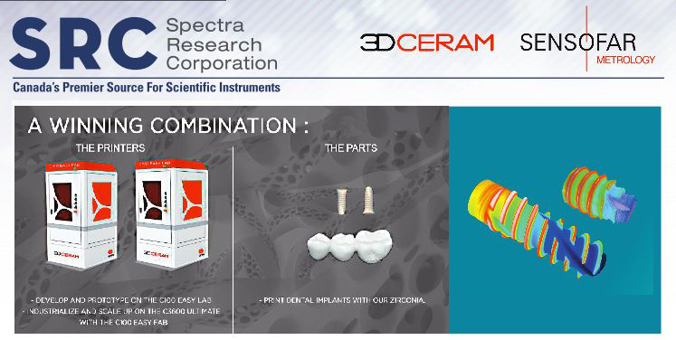
3D printing of ceramics enables a design freedom that is not achievable using tradition methods in the dental industry.
We will discuss how we can achieve with our Ceramics 3D printers tight tolerances while maintaining high strength and hardness that are
comparable to tradition manufacturing technologies.
Case studies will highlight some of the success stories that we have achieved for dental applications.
Our experts will bring you all they have learned from the multiple surface measurements they have taken on samples from the dental implant field.
Key Topics:
| Guest Speakers: | |
| Peter Durcan VP of Sales for North AmericaPeter Durcan with over 3 years in additive ceramics, a bachelor of commerce degree holder with an MBA from the Open University, United Kingdom. Peter, an Irish national who has been running the North American market with 3D Ceram for the past 2 years. |
 |
| Natalia Bermejo Product SpecialistNatalia joined Sensofar shortly after finishing her bachelor’s degree in Nanoscience and Nanotechnology at Universitat Autònoma de Barcelona (UAB) in 2018. Since then, she has been providing technical and application’s support for the Sales Team, as well as training and installing systems for prospective clients and customers. In 2020, Natalia moved to Silicon Valley, providing support to high-tech companies, as well as being the bridge between Sensofar and California’s distributor of the Sensofar brand. Currently, Natalia has returned to Barcelona HQ, and she’s has taken on an additional role supporting the Marketing Team, participating in content generation for the Sensofar channel. |
 |
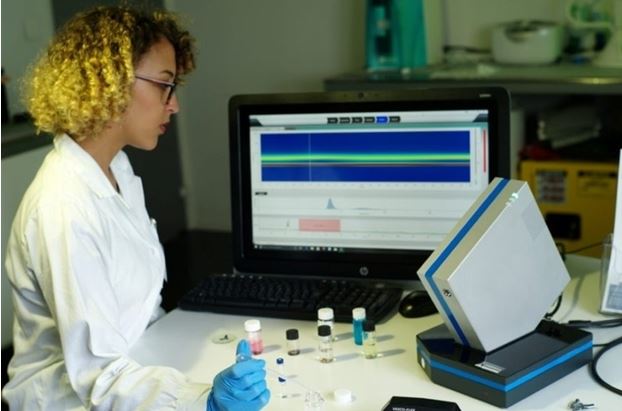
SRC is pleased to announce that we have concluded an agreement with Cordouan Technologies to be exclusive distributor in Canada of their VASCO KIN™ particle analyzer— a role we take on with great enthusiasm.
The VASCO KIN™ is a Nanoparticle Size Analyzer. A new generation of time-resolved instruments for accurate kinetic analyses combined with an in situ and contactless remote optical head. It allows for real-time monitoring of nanoparticle synthesis, agglomeration or the stability of suspensions using Dynamic Light Scattering (DLS). With a single and continuous measurement, VASCO KIN™ gives access to all characterization data of a reaction (size distribution, scattered intensity, correlogramms, etc.).
Cordouan Technologies is a French company specializing in advanced solutions for characterization (size, charge, etc.) of nanoparticles and nanomaterials. They develop, industrialize, manufacture and sell innovative instruments dedicated to academic research and industrial applications (process monitoring, quality control and R&D).
Cordouan Technologies’ unique product portfolio stems from patented and innovative technologies transferred from prestigious institutes, including Institut Français du Pétrole (IFPEN), Karlsruhe Institute of Technology (KIT) and Institut Charles Sadron (ICS).
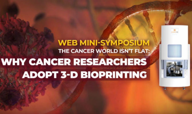
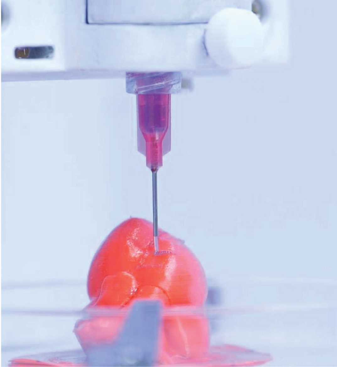
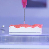
The growing push for 3-D tissue models is limited by challenges in automated handling, processing and scalability of the technology to various types of materials and high-throughput applications. To meet these challenges, ROKIT Healthcare has developed all-in-one bioprinting platforms and human-cell derived ECM bioinks that allow researchers to biologically mimic the formation of complex, heterogeneous 3-D structures and to scale the technology to high-throughput applications. This webinar will focus on the platform’s usage in cancer biology. It will discuss evidence for 3-D bioprinting capturing altered proliferation and cell morphology profiles, revealing more realistic drug responses, and mimicking vascularized tumor microenvironments – to suggest why bioprinting of cancer cells is a scientifically rigorous method to generate physiologically relevant cancer models for preclinical screening and testing, as well as for new therapy research. You will learn about:
– Key advantages of 3D bioprinting over other techniques
– Five alterations in cellular physiology of cancer cells in 3D vs. 2D
– Evaluation of how 3D bioprinted tissues have altered proliferation and cell morphology
– Evaluation of how 3D bioprinted tissues reveal more realistic drug responses
– Evaluation of how 3D bioprinted tissues mimic complex tumor-immune microenvironments
– Built-in cell incubator, diverse material use, and high-throughput capabilities of ROKIT Healthcare’s all-in-one bioprinting platform Dr. INVIVO 4D6
 |
Dr. Sanjay GuptaProfessor & Research Director
Dr. Sanjay Gupta is a Professor & Research Director and holds Carter Kissell Endowed Chair in Urologic Oncology in the Department of Urology at Case Western Reserve University and The Urology Institute at the University Hospitals Case Medical Center. Dr. Gupta’s research focuses on understanding the risk factors and mechanisms of prostatic and bladder diseases, develop appropriate biomarkers for early detection and prognosis, identify novel targets to monitor the efficacy of various synthetic and natural agents and develop them as chemo preventatives/therapeutics Dr. Gupta has authored some 150 publications, including book chapters, research articles and reviews, and has spoken at several occasions in cancer prevention symposium, seminars and meetings. He has been serving in various study sections at National Cancer Institute (NCI) and Department of Defense (DOD), and other councils around the globe. |
|
 |
Madhuri DeyPh.D Candidate @Ibrahim Ozbolat Lab – Penn State University
Talk Title: 3D BIOPRINTING OF THE CANCER MICROENVIRONMENT
|
|
 |
Da-Yae LeeSenior Bio-Consultant @ROKIT Healthcare
Talk Title: ALL-IN-ONE BIOFABRICATION IS THE KEY TO UNRAVELING CANCER COMPLEXITIES
|
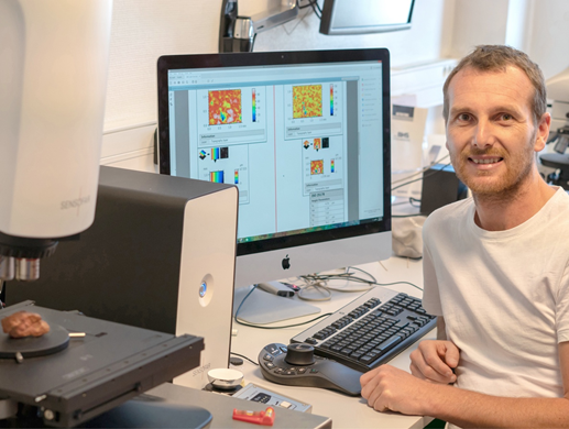
Received bachelor’s degree in Engineering Physics at Polytechnic University of Catalonia (UPC) and MSc in Nanoscience and Nanotechnology at University of Barcelona (UB). As a Sales Specialist, I’ve helped customers all around the world to choose Sensofar’s systems, no matter the field they are in. Some of my duties are to communicate Sensofar’s knowledge about optical metrology and train our customers on how to extract all the potential from our 4-in-1 technology equipment.
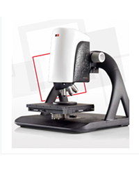
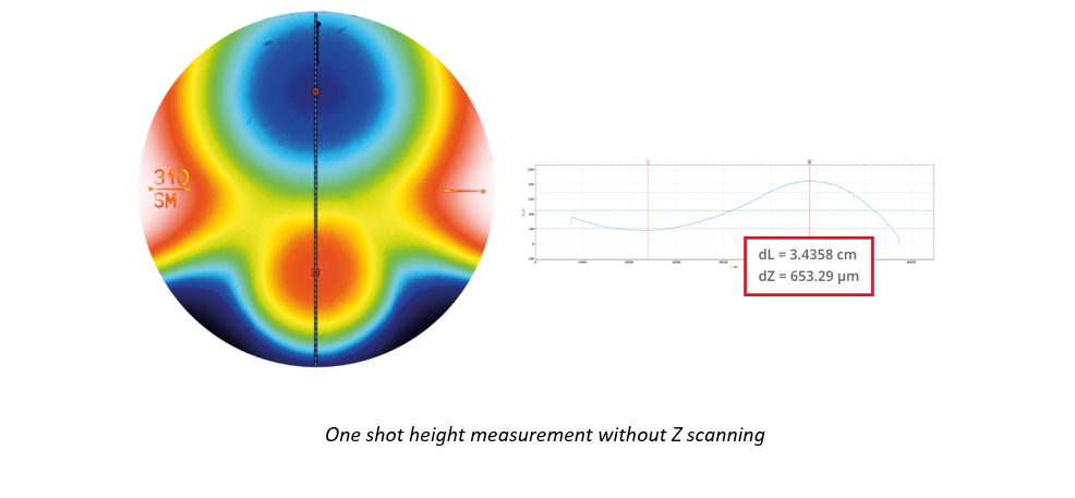
SRC is pleased to introduce Sensofar Metrology’s new metrology tool for wide areas—the S wide. The S wide is a dedicated system designed to rapidly measure large sample areas up to 300 x 300 mm. It provides all the benefits of a digital microscope integrated into a high-resolution measuring instrument. On top of that, with single button acquisition, it is extremely easy to use.
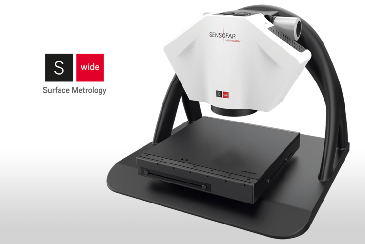
Large-area 3D optical system
The S wide is a large-area 3D optical system providing solutions in the following fields:
S wide features
Traceability
Every S wide is manufactured to deliver accurate and traceable measurements. Systems are calibrated using traceable standards according to ISO 25178 and VDI 2634-2.
Request a quote
Click here to request a quote on a Sensofar Group product.
About Sensofar Metrology
Sensofar Metrology is a member of the Sensofar Group, headquartered near Barcelona, a technology and innovation hub. Sensofar Metrology’s mission is to develop, manufacture and market high-end 3D surface metrology instruments. They also provide consultancy within the field of metrology, and pursue a philosophy of guaranteeing advanced techniques, high quality and customer service.
As we did during the month of April, we would like to share a virtual coffee with you again this coming June.
Join our Four4Free weekly webinars to hear best-in-class metrology tips from our most experienced experts. This month we are focusing on a more technical field to get the most out of optical profilers in different applications.
The live events will take place every Thursday in June, one full hour of exciting keynotes and will allow time for your questions too.
We want to bring our knowledge to meet your new ideas!
OUR FIRST APPOINTMENT
Learn more about the unlimited world of surface metrology
|
||||||||||
|
 |
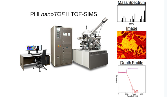 |
PHI WEBINAR SERIES:TOF-SIMS 101: Introduction, Ion Beams, MS/MS, and Materials Applications
FREE LIVE WEBINARThursday, May 14, 2020 – 10:00am (Chicago)
Time-of-Flight Secondary Ion Mass Spectrometry (TOF-SIMS) is a powerful analytical technique that can provide elemental and molecular information with high sensitivity from the sample surface to tens of microns into the sample. Additionally, TOF-SIMS can produce chemical images with high spatial resolution (<70 nm). This is achieved by utilizing a variety of ion beams to either analyze (Binq+, Au nq+, C60q+, Ga+), sputter (O2+, Cs+, Ar+, large gas cluster Ar), or mill (Ga+ FIB) the sample. The most recent advancement in TOF-SIMS is the capability of MS/MS (i.e. tandem MS) which enables confident molecular identification. The flexibility of TOF-SIMS makes it a valuable tool to investigate a wide range of materials. This webinar will introduce the fundamentals of TOF-SIMS, discuss ion beams and MS/MS, and show materials applications.
WHEN
WHAT
PRESENTER
PLEASE CLICK THE LINK BELOW TO REGISTER FOR THE EVENT! |
Sensofar Metrology is one of two divisions of the Sensofar group, based in Barcelona, Spain, an innovation and technology hub. Sensofar Metrology is renowned for its:
3D surface metrology is the measurement and characterization of micro- and nano-scale features on natural or manufactured surfaces. This is done efficiently by capturing the 3D spatial coordinates of points on a surface using a non-destructive optical technique.
Surface topography at the nanometer level
Optical surface profilers have crucial advantages over tactile approaches:
The most common optical techniques available are confocal, interferometry and focus variation, each of these has their own strengths and weaknesses.
Superior vertical resolution with confocal
Confocal: Confocal profilers measure the surface height of smooth to very rough surfaces, with spatial sampling as low as 0.10 μm—ideal for critical dimension measurements. High NA (0.95) and high magnification (150X) objectives are available to measure steep local slopes >70° on smooth surfaces with and up to 86° on rough surfaces. Sensofar’s proprietary confocal algorithms provide vertical repeatability on the nm scale.
Interferometry: White-light vertical scanning interferometers (VSI) measure the surface height of smooth to moderately rough surfaces, providing nm vertical resolution regardless of the NA. Sensofar’s new S neox optical 3D profiling microscope can use all available magnifications to profile shape features with no compromise in height resolution.
Focus variation: Focus variation has been developed for measuring the shape of large rough surfaces. Sensofar’s implementation of this approach has been specifically designed to complement confocal measurements at low magnification. Highlights of the technology include high slope surfaces (up to 86°), highest measurement speeds (mm/s) and large vertical range. This combination of features is largely suited to tooling applications.
New S neox: The new S neox optical 3D profiling microscope outperforms all previous microscopes of its kind in terms of performance, functionality, efficiency and design. It combines all three of the above techniques: confocal (best for surfaces with high slopes), interferometry (highest vertical resolution) and focus variation (measures shape in seconds). The S neox does all this in the same sensor head without any moving parts. The S neox delivers three-in-one technologies for class-leading areal measurement.
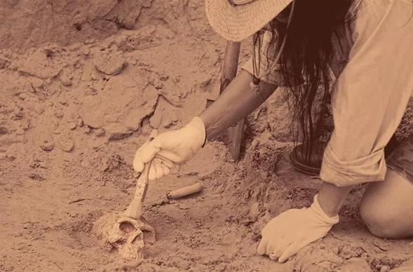
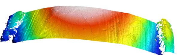
S neox helps in archaeological study of an ancient rock drawing
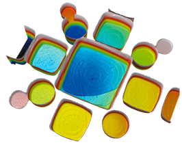
S neox Five Axis: The S neox Five Axis S measures samples at different positions of rotation and elevation,thereby generating a group of individual measurements. The SensoFIVE software merges all of the surfaces, providing a sample surface with high accuracy by using the stacked image information of each single surface measurement. Merging different elevations, the system can provide shape and form information on sharp edges and/or critical surfaces.
Request a quote
Click here to request a quote on any Sensofar product.
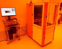
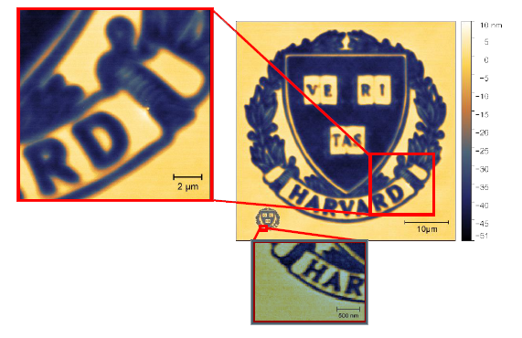
This fall SwissLitho is releasing its newest product “NanoFrazor Explore DLS“. This unique hybrid nano-micro lithography system, which combines thermal Scanning Probe Lithography (tSPL) with Direct Laser Sublimation (DLS) was developed by Heidelberg Instruments and SwissLitho.
SwissLitho commercialized tSPL out of IBM Research in 2014 and its commercial NanoFrazor systems are installed at various institutions and used for the fabrication of nanodevices when usual nanolithography techniques get complicated or fail.
You are cordially invited to this workshop which aims to introduce the capabilities of the technologies of Heidelberg Instruments and SwissLitho and discuss their opportunities for McGill University researchers.
| Program: | ||
| 1:30 pm | NanoFrazor lithography – an overview | |
| 1:55 pm |
NanoFrazor DLS – mix&match lithography in the same resist and same system | |
| 2:20 pm |
Overview on various pattern transfer processes for NanoFrazor lithography | |
| 2:45-3:00 pm |
Open user discussion | |
| 3:00-5:00 pm |
Live System demo in CR |
For more information please contact : Serge Dandache

Optical tensiometer; Smart interfacial measurement solutions for wettability and adhesion
The new generation Theta Optical Tensiometer (Attension® Theta Flex) from Biolin Scientific is a contact angle meter that enables all measurements in one instrument for both research and quality control. It measures static and dynamic contact angle, 3D surface roughness, surface free energy, surface and interfacial tension, and interfacial rheology.
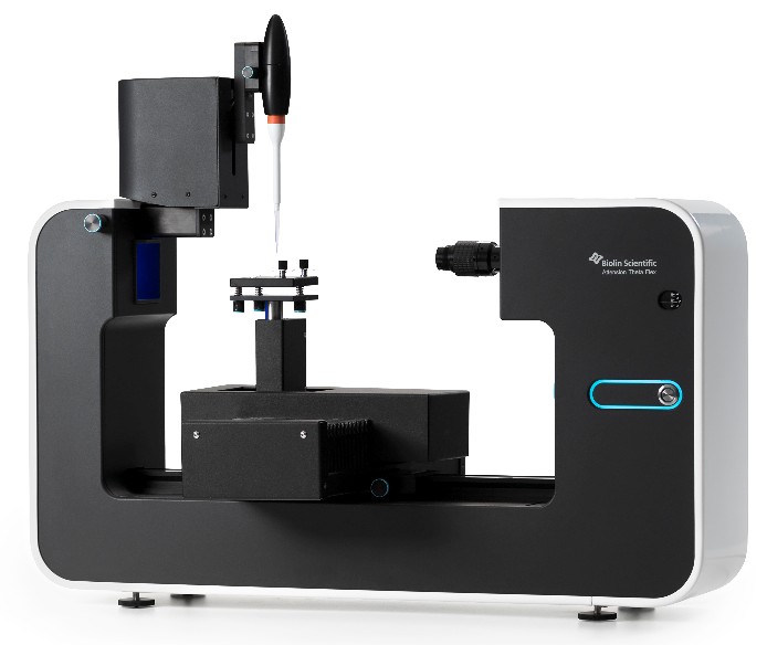
One instrument for all your measurement needs
All the measurements are readily included in the software. Thanks to the modular design, all applications can be fulfilled with one instrument and the instrument can be tailored for your needs.
Results you can rely on
High-end imaging together with sophisticated analysis algorithms detect and analyze the contact angle and surface free energy precisely. The effect of roughness to wettability can be measured with the unique 3D Topography module.
Speed and repeatability
All steps from loading the measurement to performing it and analyzing the data can be automated. The need for time consuming preparations and cleaning are removed with the disposable liquid tips.
Applications
Optical tensiometers are used in a great variety of industries and research areas, such as chemicals, pharmaceuticals, electronics, foods, energy, paper and packaging. Attension Theta Flex can be used for convenient and precise studies of:
Measurements
Attension Theta Flex can perform a complete range of measurements including:
OneAttension software
OneAttension features an intuitive user interface, live analysis and configurable user groups and accounts. In-depth analysis of your results takes a few seconds and data can easily be exported.
Modules and accessories
Attension Theta Flex enables you to choose the level of automation and the advanced functionalities that you need for your applications. With the modular design and an extensive range of modules and accessories you have room to upgrade or change the instrument as your needs evolve.
Attension Theta Flex: one instrument for all your measurement needs.
For more information or to request a quote, please contact us.