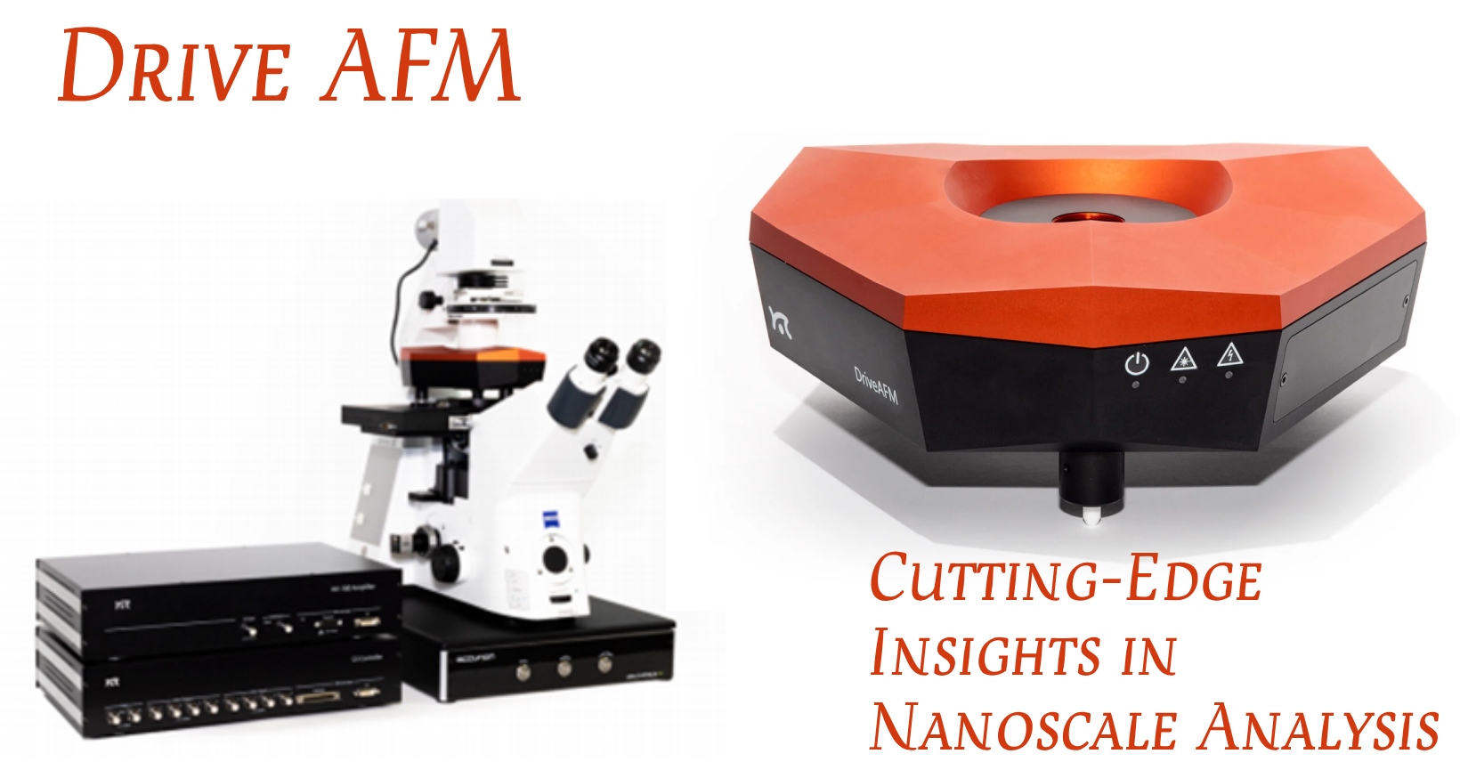
We are excited to host a two-day DriveAFM event in collaboration with Nanosurf at McGill University, Montreal. This event will feature three talks about advanced AFM techniques and applications as well as a workshop on the high-performance DriveAFM from Nanosurf.
Date: April 17-18, 2024
Location: Ernest Rutherford Physics Building, Room 103, McGill University, Montreal.
Workshop Details:
Bring a few samples, get them tested, and experience the capabilities of the DriveAFM!
The DriveAFM is Nanosurf’s novel flagship AFM platform: a tip-scanning atomic force microscope (AFM) that combines, for the first time, several capabilities in one instrument to enable novel measurements in materials and life sciences. The DriveAFM overcomes the drawbacks of other tip-scanning instruments and provides atomic resolution together with fast scanning, fast force spectroscopy, and large scan sizes up to 100 µm. Thanks to Nanosurf’s innovations in optical beam path engineering and scanner design, the DriveAFM scan head features photothermal actuation and full motorization for superior research performance and is easy to use for researchers at all levels of experience.
- CleanDrive: stable excitation in air and liquid
- Ultra-low noise
- Direct drive: high-resolution imaging and large scan area
- Fully motorized system: full control via software
Agenda:
April 17th, 2024 (Wednesday):

April 18th, 2024 (Thursday):

Speaker Details:

Speaker Name and Title: Prof. Peter Grütter, Scientific Director and Founder of the McGill Nanotools Microfabrication Facility Talk Title: Interpreting tapping (AC) operation mode Talk Summary: Tapping or AC mode is often used in AFM imaging leading to high-quality images. Interpreting the measured images is often challenging, as the contrast depends on the operation parameters and how the AFM is set up. In this short talk, I will discuss the relevant theory behind tapping and the practical aspects of what to watch out for to facilitate the interpretation of the acquired data. |

Speaker Name and Title: Prof. Angelo Gaitas, Assistant Professor (Icahn School of Medicine at Mount Sinai) Talk Title: Advancements in Fluid Micro Cantilevers and Novel Thermocouple Devices for Tissue and Cellular Analysis Talk Summary: This talk will cover developments in bioAFM innovations in my laboratory. First, we utilize fluid micro cantilevers in atomic force microscopy (AFM) for a range of applications, including the precise measurement of single-cell mass in a media environment. This advanced technique allows for a detailed analysis of the nano-mechanical properties of human induced pluripotent stem cells (iPSCs) and their differentiation into cardiomyocytes (iPSC-CMs). The employment of fluid micro cantilevers in AFM enhances the accuracy and scope of our measurements, revealing significant changes in cell elasticity and mass during iPSC differentiation. These findings establish elasticity and mass as key indicators in evaluating the development of iPSCs, providing invaluable insights for cell therapy, drug testing, and cardiac disease research. Our study demonstrates the capability of AFM, especially with the use of fluid micro cantilevers, to effectively differentiate cells pre- and post-differentiation based on their mechanical properties. This advancement underscores the potential of these techniques as morphological markers in iPSC research. The results, while promising, necessitate further studies to confirm their generalizability to other cell lines. Additionally, our work points to the necessity of developing more refined AFM measurement techniques in fluid media, proposing various methods to enhance the technology’s resolution and accuracy in future research applications. Second, we have developed a novel thermocouple device tailored for intracellular temperature measurement. Temperature regulation and gradients are crucial in biological research, as thermal events significantly impact cellular functions. This microcantilever thermocouple sensor combines doped silicon and gold to form a sensitive junction, suitable for biological applications. Its design ensures mechanical robustness, high sensitivity, and rapid response, ideal for liquid environments and minimal impact on cellular processes. The fabrication involves several precise steps, resulting in a sensor with a high Seebeck coefficient (447 μV/°C) and millisecond response time. This advanced device has demonstrated effective and precise transient thermometry in biological samples, showing its potential in understanding and measuring thermal events at a cellular level. |

Speaker Name and Title: Dr. Edward Nelson, Applications Scientist, Nanosurf Talk Title: Photothermal Torsional Resonance Imaging for 2D Materials Characterization Talk Summary: Stacked layers of 2D materials such as graphene and hBn show remarkable electrical, optical and magnetic properties depending on the angle between the layers. A common approach to measure this angle is to use an Atomic Force Microscopy (AFM) to visualize the Moiré superlattice that forms from interactions between the layers. Piezoresponse Force Microscopy (PFM), a type of imaging mode, offers high contrast but is only effective when the sample/substrate is conductive. Regrettably, this method is not effective when the sample is on a non-conductive substrate, like a transfer polymer. Torsional Resonance Microscopy (TRM) is a method to drive the torsional resonances of the AFM cantilever and has shown remarkable contrast of the Moiré superlattice. In addition, because the cantilever is driven mechanically, the method works on all types of substrates. Unlike piezo-acoustic TRM, photothermal TRM does not require any special hardware outside of what is already available on the DriveAFM. In addition, because the cantilever is driven photothermally, it can work under liquids without introducing parasitic coupling with the environment. This new mode is expected to open up new research opportunities for materials characterization. |
Register for Free using the form below.


