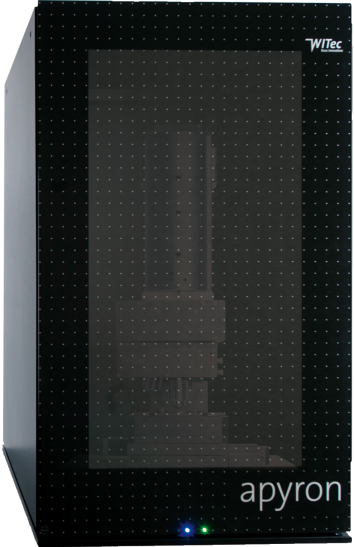
With the apyron imaging system you will be prepared to explore beyond the established frontiers of your field. It has been developed to overcome the boundary between ease-of-use and ultimate capability in Raman Imaging. Equipped with this seamless versatility, your creativity can be fully expressed.
When simplicity meets performance
As a member of the WITec product family, apyron sets the benchmark for automated Raman Imaging systems with excellent imaging qualities: outstanding spectral and spatial resolution, ultra-fast acquisition times, and exceptional signal sensitivity in combination with automated system configurations and intuitive measurement procedures.
When high-resolution imaging meets high-resolution spectroscopy
Ultrahigh-throughput spectrometers provide unprecedented spectral resolution, detailed spectral information at every image pixel and the highest Raman signal sensitivity.
apyron is the ideal Raman imaging system for:
- Multi-user labs accomodating varied user skills and requirements
- Industry labs with recurring experimental scenarios and an emphasis on time-critical turnover
- Raman newcomers with advanced imaging requirements
- Leading Raman spectroscopists seeking the next performance benchmark
The new automated Raman Imaging system apyron
A New Class of Automated Raman Imaging Systems – Key Features
WITec is established as the manufacturer of the industry‘s most advanced Raman Imaging systems. This distinction now also extends to automated systems.
TruePower: Automated Absolute Laser Power Determination in mW
The absolute laser power is measured in the optical fiber and can be adjusted with accuracy of <0.1 mW. A laser shutter shields the sample from the laser light and opens only during Raman analysis with optimized laser power to avoid any sample degradation. apyron is the only Raman Imaging system on the market to provide such an accuracy ensuring optimal laser power for the preservation of delicate samples and reproducibility in measurement conditions.
Automated Laser Wavelength Selection and Spectrometer Adjustment
The selection of the laser has never been easier: With a simple click of the mouse the wavelength can be chosen in the software user interface. What follows is an automated adjustment and calibration of all spectrometer components such as filters, gratings etc. to enable optimal system performance.
Performance Facts:
- More than 16 million Raman spectra can be acquired in a single dataset
- Spectral resolution down to 0.1 rel. 1/cm per pixel (@633 nm excitation)
- 3D Raman Imaging at a lateral resolution limited only by physical law
- Push-button instrument and measurement control
Ultrahigh-throughput Spectrometers for Unprecedented Spectral Resolution, Detailed Spectral Information at Every Image Pixel and the Highest Raman Signal Sensitivity
The lens-based imaging spectrometer was specifically designed for Raman microscopy and applications at ultra-low light intensities. Versions are available for a wide variety of excitation wavelengths and focal lengths and are optimized for their specific laser wavelength:
- UHTS 300 for most effective and ultrafast 3D confocal Raman Imaging
- UHTS 400 for excellent spectral and image quality for the red and NIR
- UHTS 600 for unrivaled spectral resolution in 3D confocal Raman Imaging
Additional Features:
- Automated sample positioning and motorized scan tables
- Software-controlled measurement settings
- TrueSignal: system-controlled adjustment of the optical fiber position to maximize the light into the fiber output of the microscope
- TrueCal: convenient performance of pre-defined calibration routines for the optical and mechanical microscope components whenever required
- Laser safety class 1
apyron: powerful data acquisition and post-processing
apyron Applications
Carbon Tetrachloride in an Alkane-Water-Oil Emulsion
Carbon Tetrachloride is quite often used as a reference sample in order to determine the performance of a Raman spectrometer in terms of spectral resolution. The characteristic peak at 460 rel. 1/cm should be clearly resolvable at room temperature. Due to its ultra-high optical throughput the apyron is the first system on the market to allow for FAST RAMAN IMAGINGTM while simultaneously maintaining the ability to resolve this peak.

3D confocal Raman image of the emulsion. Green: Alkane; Blue: Water; Yellow: CCl4 + Oil. (Image parameters: 200 x 200 x 20 pixels, 100 x 100 x 10 µm³ scan range, 0.06 s integration time per spectrum, 532 nm excitation wavelength.) Zoom-in image with high spectral resolution. (Image parameters: 100 x 100 pixels, 10 x 10 µm², 0.08 s integration time per spectrum, UHTS 600 spectrometer, 1800 g/mm grating.) Due to the high spectral resolution of the spectroscopic system the bands of the CCl4 peak at 460 cm-1 can be clearly resolved at room temperature.
White-light microscopy and color-coded Raman Image overlay of a squashed banana pulp sample
The Raman image was acquired with an apyron attached to the 600 mm focal length UHTS 600 spectrometer system (grating: 300 g/mm) to provide the highest spectral performance. Red: beta-Carotene-rich areas, Green: Starch, Blue: Water

Only the apyron is capable of generating Raman images along with the parameters as in the following: Excitation: 532 nm, Laser Power: 49.9 mW, Scan area: 400 x 300 µm², 1200 x 900 pixels, Integration time: 2 ms/spectrum/pixel


