Reichert Quartz Window Flow Cell

Reichert Quartz Window Flow Cell

  • Surface plasmon field enhanced fluorescence spectroscopy
  • Extremely sensitive and effective tool for detecting and quantifying biomolecular binding
  • Left and right fluid path imaged after binding FITC labeled antibody to sensor slide surface.
  • features a quartz window in each of the fluidic pathways to enable SPR experiments combined with Photochemistry/ Fluorescence.


SKU: Quartz Window Flow Cell Category:


Description

Reichert Systems

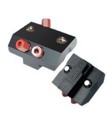
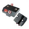
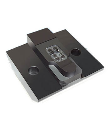
The Quartz Window flow cell features a quartz window in each of the fluidic pathways to enable SPR experiments combined with Photochemistry/ Fluorescence.

The Reichert quartz window flow cell facilitates experiments combining SPR with photochemistry, imaging fluorescent labeled molecules on the sensor surface by direct excitation and surface plasmon field enhanced fluorescence spectroscopy. Surface plasmon field enhanced fluorescence spectroscopy is an extremely sensitive and effective tool for detecting and quantifying biomolecular binding. This technique depends on excitation of a fluorophore near the gold sensor surface of an evanescent field. Resonance of p-polarized light with surface plasmons (oscillating electrons) in the gold layer produces the evanescent field.

Utilizing the Quartz Flow Cell


Figure 1. Picture of microscope over SPR Instrument. Transmitted light fluorescent microscope imaging. Shown is the quartz window flow cell mounted on the Reichert Technologies SR7000DC surface plasmon resonance spectrometer.

https://asoft8280.accrisoft.com/reichert/clientuploads/directory/FIG-2-QUARTZ CELL.jpg
Figure 2. Quartz Window Flow Cell – Top View

https://asoft8280.accrisoft.com/reichert/clientuploads/directory/FIG-3-QUARTZ CELL.jpg
Figure 3. Left and right flow paths imaged while flowing an FITC labeled antibody over the left channel surface. FITC label excited by light from above the sensor surface (through buffer).

https://asoft8280.accrisoft.com/reichert/clientuploads/directory/FIG-4-QUARTZ.jpg
Figure 4. Left and right fluid path imaged after binding FITC labeled antibody to sensor slide surface.